
TERTp methylation was positively correlated with chromatin accessibility and TERT mRNA expression in 8 melanoma cell lines. In spite of the high promoter methylation fraction in wild-type (WT) samples, TERT mRNA was only expressed in the melanoma cell lines with either high methylation or intermediate methylation in combination with TERT mutations.
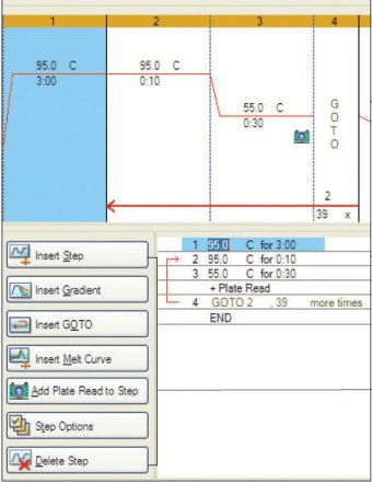
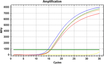
#Bio rad cfx manager 3.1 normalize skin
We unexpectedly observed that the methylation of TERTp was as high in a subset of healthy skin cells, mainly keratinocytes, as in cutaneous melanoma cell lines. This study aimed to elucidate the combined contribution of epigenetic (promoter methylation and chromatin accessibility) and genetic (promoter mutations) mechanisms in regulating TERT gene expression in healthy skin samples and in melanoma cell lines (n = 61). Furthermore, two hotspot mutations in TERTp, dubbed C228T and C250T, have been revealed to facilitate binding of transcription factor ETS/TCF and subsequent TERT expression. CpG methylation in the TERT promoter ( TERTp) was correlated with TERT mRNA expression. γ value was changed to 0.The telomerase reverse transcriptase ( TERT) gene is responsible for telomere maintenance in germline and stem cells, and is re-expressed in 90% of human cancers. Numbers of sox17-positive cells seen in dorsal view and standard deviation are given (upper right) as well as number of analyzed embryos ( n) (lower right). FHD-GFP lead to complete loss of forerunner cells (black arrow) in the majority of MZ sur embryos ( p, t see also Additional file : Statistical analysis of forerunner cells). Number of forerunner cells (black arrow) is reduced in MZ surcontrol and after MOs injections ( o, q, s).

m– t sox17 in-situ hybridizations show only slight reduction of endoderm after injection of MOs ( q– t). Percentage of embryos showing the same phenotype as in the image is given (upper right). Co-injection of FHD-GFP and MOs also results in a decreased staining ( j, l) when compared to FHD- GFP injection ( f, h).

dre-miR-430 morpholinos (MOs) massively reduce axial foxa2 signals in both genotypes ( l, k). Injection of FHD-GFP causes a broadened axial signal in 50% of wild type embryos, but no reduction of axial cells ( f), and strengthens the effect in 60% of the MZ sur mutants ( h). e– l foxa2 in-situ hybridizations show reduction of axial mesoderm formation in MZ surmutants. Injection of FHD-GFP in MZ sur mutants enhances the phenotype ( d/ d′ note reduced size and additionally discontinuity of staining). In uninjected MZ sur embryos the width is reduced ( c′). col2a1a in-situ staining in wild-type (un-injected control ( a) or injected with FHD-GFP ( b)) shows wild-type notochord of expected width (white brackets in enlarged sections a′ and b′). a– dWild-type ( a, b) and MZ sur mutant ( c, d) embryos at 24hpf. FHD-GFP interferes with severity of the MZ sur mutant phenotype.


 0 kommentar(er)
